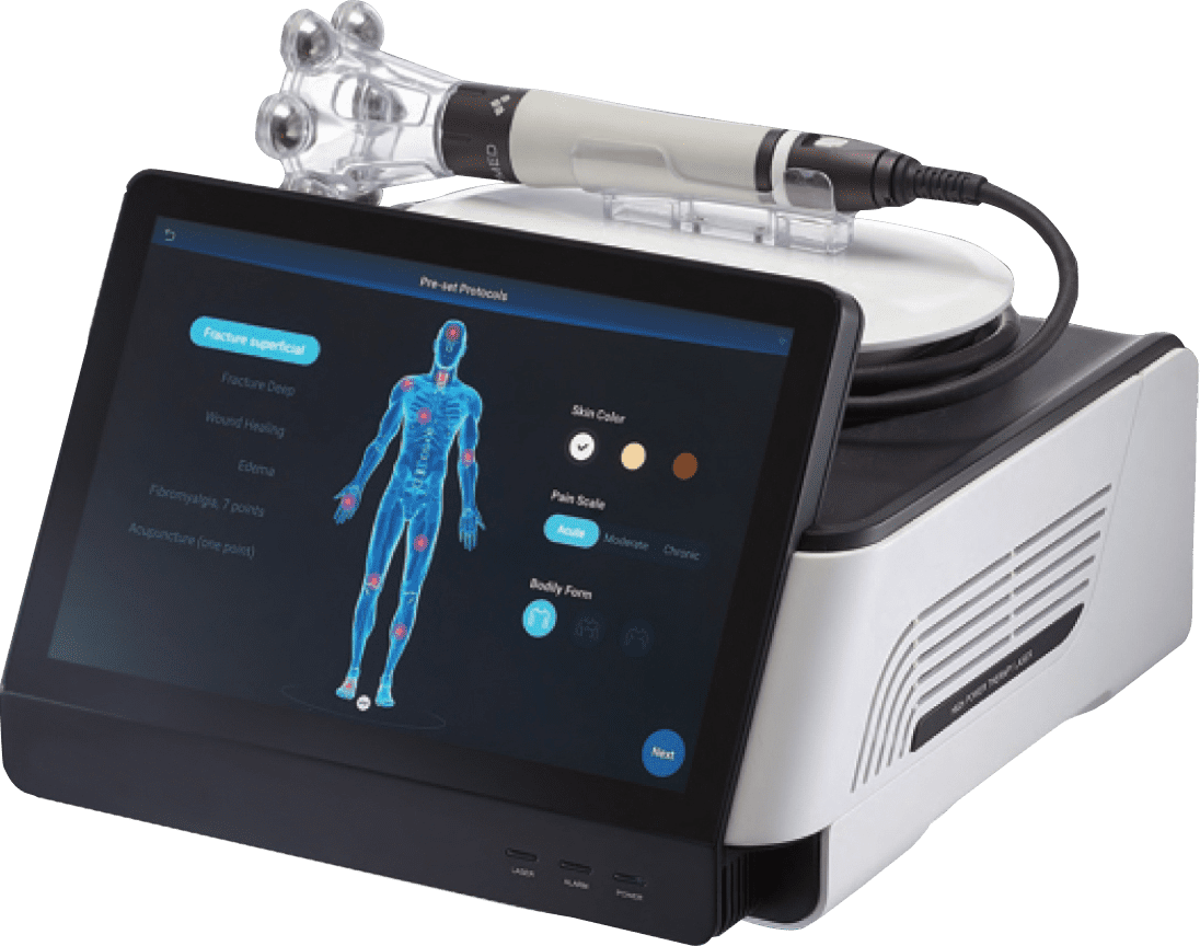
CLASS IV THERAPY LASERS
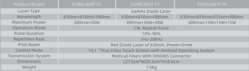
TECHNICAL PARAMETER
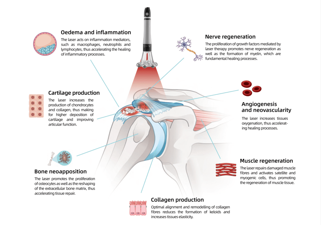
EFFECTS ON METABOLIC PROCESSES
PHOMED Laser Class IV therapeutic lasers deliver specific red and near-infrared wavelengths of laser light to
induce a photochemical reaction and therapeutic effect. Physiological effects include increased circulation,
reduced inflammation, pain reduction and enhanced tissue healing.
During each painless treatment, laser energy
increases circulation, drawing water, oxygen, and
nutrients to the damaged area. This creates an
optimal healing environment that reduces inflam-
mation, swelling, muscle spasms, stiffness, and
pain. As the injured area returns to normal, function
is restored and pain is relieved.
During PHOMED Laser Therapy, infrared laser light
interacts with tissues at the cellular level increasing meta-
bolic activity within the cell. By improving the transport of
nutrients across the cell membrane, the increased
production of cellular energy (ATP) is stimulated. The
cascade of beneficial effects that follows includes
increased cellular function and tissue repair.

WHY ARE WAVELENGTHS IMPORTANT?
Wavelengths determine laser energy’s depth of penetration in tissue and specifically target beneficial chro-
mophores to stimulate the photochemical reaction.
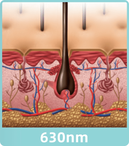
Irradiating an area with 630nm,
a wavelength where melanin in
our skin absorbs very well, will
ensure a large dose to the
superficial region. Since light
can both inhibit bacteria and
promote cell growth, laser
therapy has incredible results in
wound healing and scar tissue
regulation.
The quicker oxygen is released
into the blood stream, the more
fuel the cell has to carry out all
of its natural healing process-
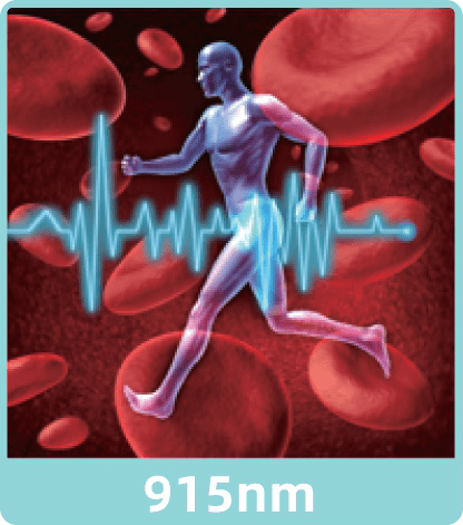
es. The peak of hemoglobin’s
absorption lies at 905nm, and
when this radiation is absorbed,
more of this oxygen-fuelis made
available to the cells.
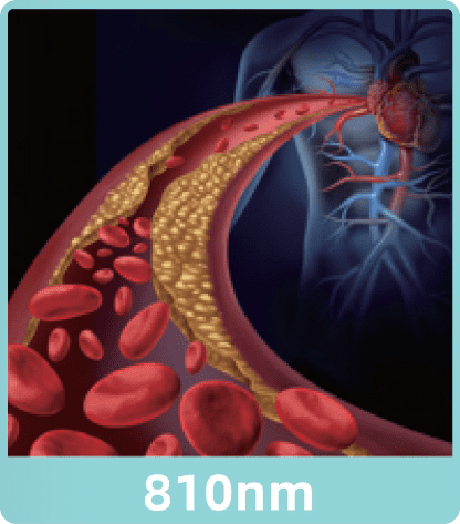
The enzyme determining how
efficiently the cell converts molec-
ular oxygen into ATP has the
highest absorption at 810nm.
Regardless of the enzyme’s molec-
ular state, when it absorbs a
photon it will flip states. Photon
absorption will accelerate the
process and increase cellular ATP
production.
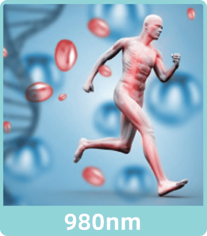
Water in the blood transports
oxygen to the cells, carries waste
away, and absorbs very well at
980nm. The energy created from
absorbing a photon gets convert-
ed to heat, creating a temperature
gradient at the cellular level,
stimulating micro-circulation, and
bringing more oxygen-fuel to the
cells.
Precise Boundary
- The treatment planning software enables conformal ablation of the whole tumor with no
upper limit on the volume nor tumor shaPe - The margin between treated and untreated tissue can be as narrow as 6 to 10 cells wide
Precise Dosage
- Real-time imaging allows visual feedback during treatment process
- Immediate image after each exposure can be compared with the previous
- An operator can adjust the dose anytime to suit the individual needs
- Integrated dose data will be recorded for future analysis and effect evaluation
Precise Control
- With +1 mm accumulative error, the accurate
movement of 6-dimensional motion system
can ablate tumors adjacent to major vessels
and nerves safely - Color Doppler Ultrasound provides clear real-time monitoring during the whole treatment
Indications
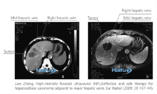
Solid tumors of soft tissues, such as liver tumor, uterine fibroids, Breast cancer, kidney tumor, pancreas tumor, bone tumor.
Contraindications
- Tumors in air-containing viscera, such as lung, stomach, and bowel
- Mediastinal tumors
- Spinal tumors
Clinical Advantages
- Noninvasive treatment to preserve
organs and structures, with no blood transfusion and no radiation - Conformal and precise ablation
- One time treatment, no limitation
by tumor size and shape - Real-time ultrasound-quided therapy
with digital quantitative analysis - Activation of immune system
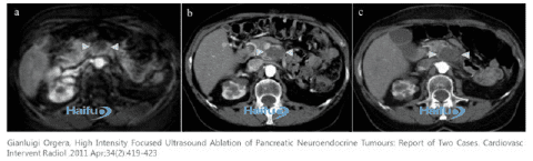
Pancreatic cancer
A 43-year-old woman with pancreatic tumor. (a) MRI before
HIFU shows a 2.4-cm lesion (arrowheads). (b) MDCT
performed 24h after HIFU shows ablation of entire lesion
(arrowheads). (c) MDCT scan 9 months after treatment
shows the lesion appears slightly diminished in dimensions
but widely hypodense and without residual enhancing
region (arrowheads).
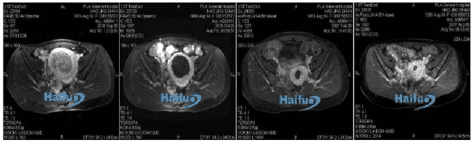
Uterine fibroids
Enhanced MRI images in a 37-year-old woman with uterine fibroids. Before treatment, the fibroid had abundant and active blood supply. Fourteen days after treatment, the fibroid showed obviously shrinkage and lost activity. MRI examination 4.5 months and 10 months after HIFU showed obvious shrinkage of treated fibroids.
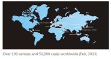
- European Institute of Oncology, Milan, Italy
- The John Radcliffe Hospital, Oxford, UK
- University Clinics of Bonn, Bonn, Germany
- Hospital Mutua de Terrassa, Barcelona, Spain
- Saint Marina Hospital, Pleven, Bulgaria
- Medical Center of Central Bank of Russian Federation,
Moscow, Russia - Queen Mary Hospital of the University of Hong Kong,
China - King Fahad Medical City, Riyadh, Saudi Arabia
- Incheon Christian Hospital, Incheon, Korea
- CIMEQ (Centro de Investigaciones Medico Quirurgicas),
Havana, Cuba - 301 PLA General Hospital, Beijing, China etc.
Professional Equipment
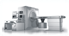
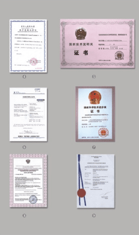
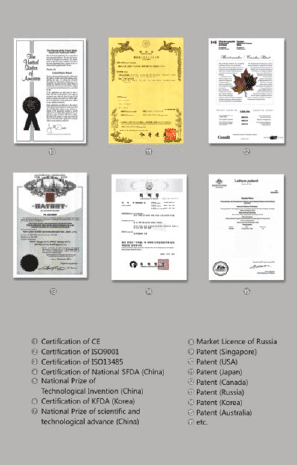
With complete intellectual rights and CE- mark it is the world first equipment clinically applied in tumor treatment a result of over 20 years experience and optimization
Experienced Specialists
A team of experienced medical and engineering specialists will provide integrated training and service to enable the end-users independent operation of the equipment.
Customized Solution
Suitable clinical protocols, operation & management advices and research cooperation proposal will be tailored for
each end-user.
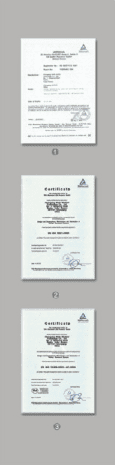
QUALIFICATION